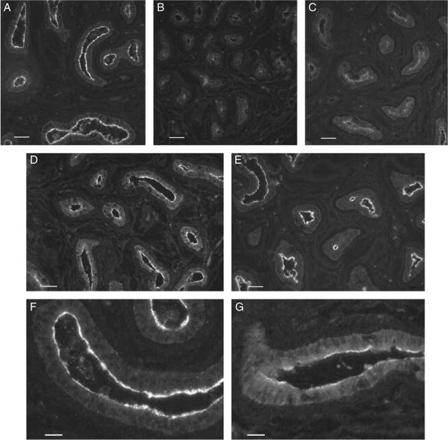
Figure 2.

AQP9 immunostaining in the cauda epididymidis of 25-day-old rats after neonatal treatment with (A) vehicle (control); (B) GNRHa; (C) DES; (D) EE; and (E) DES+TE. (F and G) Higher magnification pictures of control and EE-treated animals respectively. GNRHa and DES treatments caused a marked decrease in apical AQP9 staining. EE-treated animals showed a moderate decrease in AQP9 immunofluorescence labeling. Administration of TE maintained AQP9 expression in DES-treated rats at control levels. Digital images were acquired using identical parameters. (A–E) Bars=40 μm. (F and G) Bars=15 μm.
