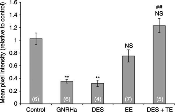
Figure 3.

Quantification of AQP9-associated fluorescence staining in the cauda epididymidis of 25-day-old control and treated rats (groups represented in Fig. 1). Significant decreases in AQP9 staining intensity were induced by GNRHa and DES. EE also induced a reduction in AQP9 staining but the difference was not significant versus control. Co-administration of testosterone with DES completely restored AQP9 staining compared to DES alone, with AQP9 staining intensity comparable to control. Data are expressed relative to control, and represent the means±S.E.M. obtained from 4 to 7 animals per group. **P<0.0005 versus control; ##P<0.005 versus DES; NS, not significant versus control.
