
Figure 5.
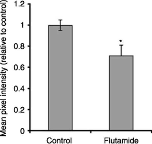
Quantification of AQP9-associated fluorescence staining in the cauda epididymidis of control and flutamide-treated 25-day-old rats. The mean pixel intensity of AQP9-associated fluorescence in the apical pole of principal cells was markedly decreased following flutamide treatment. Data are expressed relative to control and represent the means±S.E.M. obtained from three animals per group. *P<0.05 versus control.