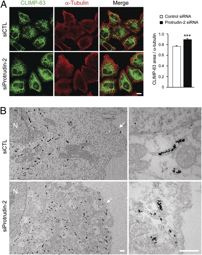
Fig. 4.
Protrudin depletion modulates the ER sheet-to-tubule balance. (A) HeLa cells were transfected with control (siCTL) (n = 33) or protrudin (n = 22) siRNAs for 72 h and then immunostained with CLIMP-63 (green) and α-tubulin (red). All studies were done at the same time, with the same procedures and microscope settings. Pixel areas of CLIMP-63 were normalized to those of α-tubulin using ImageJ tools. Data are presented in arbitrary units. Means ± SEM are shown. Student t test: ***P < 0.001. (Scale bar: 10 μm.) (B) siRNA-transfected HeLa cells were immunogold-labeled for a luminal epitope of CLIMP-63, with visualization by electron microscopy. N, nucleus. Arrowheads denote the plasma membrane. (Scale bars: 500 nm.)

