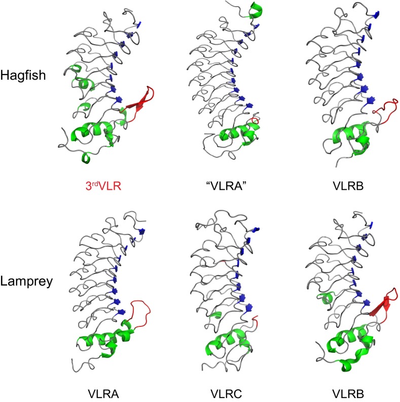
Fig. 4.
Comparison of the predicted 3D structure of hagfish 3rdVLR (GenBank accession no. KF314090) and other agnathan VLRs. The crystal structures of hagfish “VLRA” (PDB ID: 2O6Q), hagfish VLRB (PDB ID: 2O6S), lamprey VLRA (PDB ID: 3M18), and lamprey VLRB (PDB ID: 3E6J) were retrieved from Protein Data Bank (PDB). The 3D structure of lamprey VLRC (GenBank accession no. KC244064) is also predicted for comparison. β-Sheet and α-helix structures are shown in blue and green colors, respectively. Loops located in the LRRCT portion are indicated in red. Note that the hagfish 3rdVLR, lamprey VLRA, hagfish VLRB, and lamprey VLRB all possess a protruding loop in the LRRCT region, whereas hagfish “VLRA” and lamprey VLRC do not.

