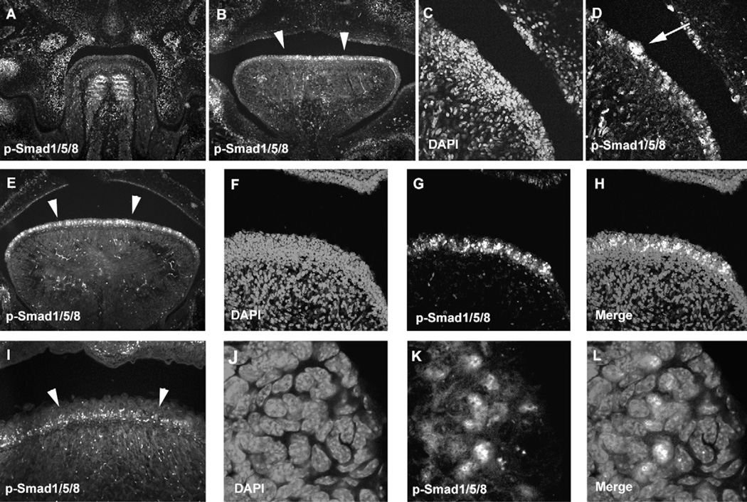
Fig. 2.
P-Smad1/5/8 in filiform papilla development. Immunohistochemistry of p-Smad1/5/8 in frontal head sections of the developing tongue at E13.5 (A), E14.5 (B–D), E16.5 (E–H, J–L) and birth (I). Arrowheads indicating p-Smad1/5/8 positive cells in tongue papillae (B, E, I). Arrow indicating p-Smad1/5/8 positive cells in fungiform papillae (D). DAPI (C, F, J), p-Smad1/5/8 (A, B, D, E, G, I, K), Merge images (H, L). (F–H, J–L) High magnification of E.

