
Figure 2.
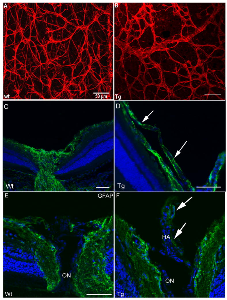
Analysis of retinas from transgenic mice overexpressing Nuc1 mutant βA3/A1-crystallin in astrocytes, and wild type littermates. Panels A and B show confocal microscopy of P21 retinal flat mounts with astrocytes immunolabelled with anti- GFAP (red). The left image (A) is from a wild type littermate. The astrocytes show a normal compact stellate structure producing a typical honeycomb-like network. In contrast, the image on the right (B), from a transgenic overexpressing the mutant (Nuc1) βA3/A1-crystallin in astrocytes, shows bundle-like structures with abnormal patterning and short, thickened processes. In panels C-F, frozen sections from eyes of wild type and transgenic mice are shown. An additional layer of astrocytes (GFAP stained green) from the hyaloid artery along the surface of the nerve fiber layer is present in transgenic mice (D, arrows), but not present in wild type mice (C). Panels E and F show GFAP (green) immunostaining of the optic nerve head from P49 wild type and transgenic mice. In wild type optic nerve head (E), GFAP-positive astrocytes are seen on the surface and inside the optic nerve head. In the transgenic mouse expressing the mutant protein (F), GFAP-positive astrocytes are present on the optic nerve head and on the surface of the retained hyaloid artery (HA, arrows). Scale bar=50 μm, n=7 wild type, 7 transgenic mutant mice.