
Figure 1.
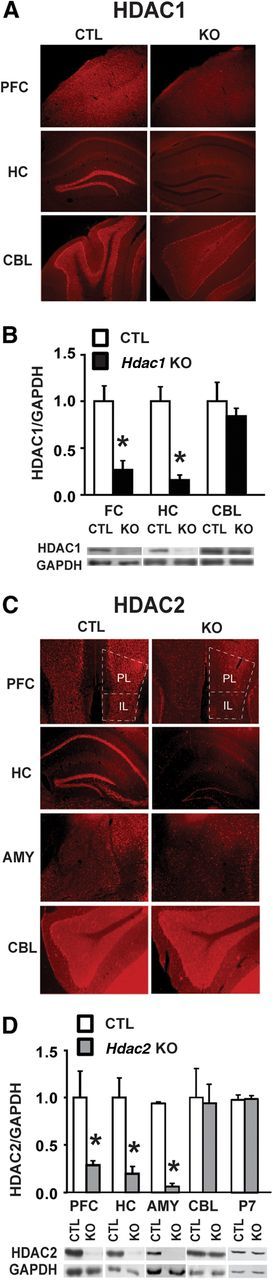
Postnatal forebrain deletion of Hdac1 or Hdac2. A, Immunohistochemistry of coronal sections of 8-week-old mouse brain demonstrates a loss of HDAC1 protein in CaMKII-Cre93-mediated conditional KO mice relative to littermate CTL mice. Shown are sections of frontal cortex (FC), hippocampus (HC), and cerebellum (CBL). HDAC1 expression was unchanged in CBL, indicative of a forebrain-specific KO. B, Western blot analysis confirmed knockdown of HDAC1 in FC and HC to 20–30% of CTL, but not in CBL. C, Immunohistochemistry images from coronal sections of 8-week-old Hdac2 KO and CTL mice demonstrating loss of HDAC2 protein in PFC, including both prelimbic (PL) and infralimbic (IL) cortex, HC, and amygdala (AMY) but not in the CBL. D, Western blot analysis confirmed a significant reduction of HDAC2 protein in PFC, HC, and AMY (∼70–90%) in the conditional KO mice compared with CTL, with no change in CBL. Forebrain levels of HDAC2 protein remained unchanged at postnatal day 7 (P7) in conditional Hdac2 KO mice compared with CTL, confirming a postnatal deletion by our CaMKII-Cre strategy. *p < 0.05.