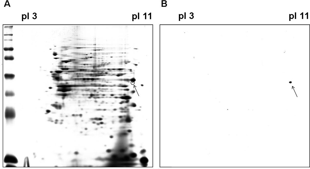
Fig. 3.
Two-DE and immunoblot analysis of SEC1-stimulated bovine PBMCs. Cell lysates from bovine PBMCs cultured with SEC1 for 8 d were resolved by 2-DE in duplicate. One gel was stained with Coomassie Blue (A) and the other was transferred to a PVDF membrane and probed with the FOXP20A mAb (B). Arrows indicate the corresponding spot in both gels.

