
Fig. 3.
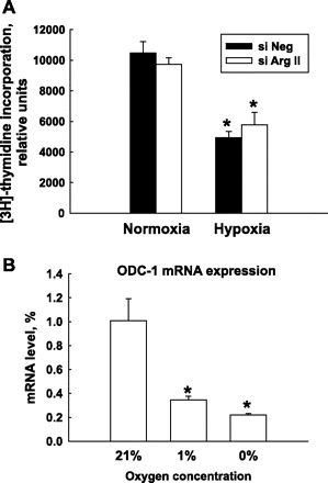
HMVEC proliferation under normoxia and hypoxia conditions. A: HMVEC were transfected with siNeg or Arg II siRNA (siArg II). Twenty-four hours after transfection, cells were exposed to 21% or 1% O2 for an additional 48 h. During the last 6 h of the exposure, [3H]thymidine was added to cells. Exposure of HMVEC to 1% O2 dramatically decreased the rate of HMVEC proliferation. Silencing of Arg II did not have an effect on HMVEC proliferation. B: expression of ornithine decarboxylase 1 (ODC-1) mRNA in HMVEC exposed to different oxygen concentrations for 24 h. The results shown are representative of three or more independent experiments made in triplicate. *P < 0.01 vs. normoxia.