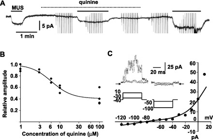
Fig. 2.

Quinine-insensitive component in MUS-induced current in guinea-pig AM cells. A: reversible suppression of 10 μM MUS-induced current by 100 μM quinine. Whole cell current was recorded at −60 mV with the nystatin method. Chemicals were added to saline during the indicated periods (bars for MUS; dotted line for quinine). B: summary of inhibition of 10 μM muscarine-induced currents by quinine of various concentrations in 4 cells. Peak amplitudes of muscarine-induced currents in the presence of quinine were expressed relative to averaged amplitudes of the currents before and after addition of quinine. These relative values were plotted against concentrations of quinine. Line represents y = 0.4 + 0.6/[1 + (x/6.1)1.7] (see materials and methods). C: current-voltage (I-V) relationship for muscarine-induced current in the presence of 100 μM quinine; 50-ms pulses were applied from the holding potential of −40 mV in 10-mV steps before, during, and after addition of muscarine to 100 μM quinine-containing saline. Muscarine-sensitive currents were obtained by subtracting current responses during addition of muscarine from averages of those before and after it. Current level at the end of pulse was measured and plotted against the command potential. Inset: muscarine-sensitive currents (top) obtained with the command pulses (bottom) in the presence of 100 μM quinine. Arrows indicate the zero current level.
