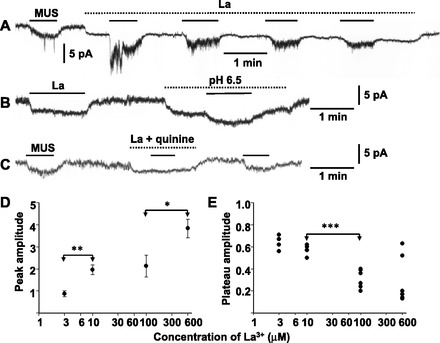
Fig. 4.

Dual action of La3+ on muscarine-induced current in AM cells. A: effects of 600 μM La3+ on 10 μM MUS-induced current. B: La3+-induced inward current is markedly suppressed by a decrease in external pH to 6.5. C: MUS fails to induce an inward current in the presence of 600 μM La3+ and 100 μM quinine. Whole cell current was recorded at −60 mV using the nystatin method in three different guinea-pig AM cells. MUS and other chemicals were added to saline during the indicated periods (bars for MUS; dotted lines for other chemicals). D: peak amplitudes of inward currents induced by first application of 10 μM MUS in the presence of La3+ are plotted against concentrations of La3+. Peak amplitude was expressed relative to that of the MUS-induced current before application of La3+ in the same cells. Data represent means ± SE of 3–4 cells. E: plateau levels of inward currents induced by third or fourth application of 10 μM MUS in the presence of La3+ are plotted against concentrations of La3+. Plateau level was expressed relative to that of the MUS-induced current before application of La3+. Peak and plateau levels were determined at the middle of current fluctuation. *P < 0.05, **P < 0.01, and ***P < 0.001, statistically significant difference.
