
Figure 1.
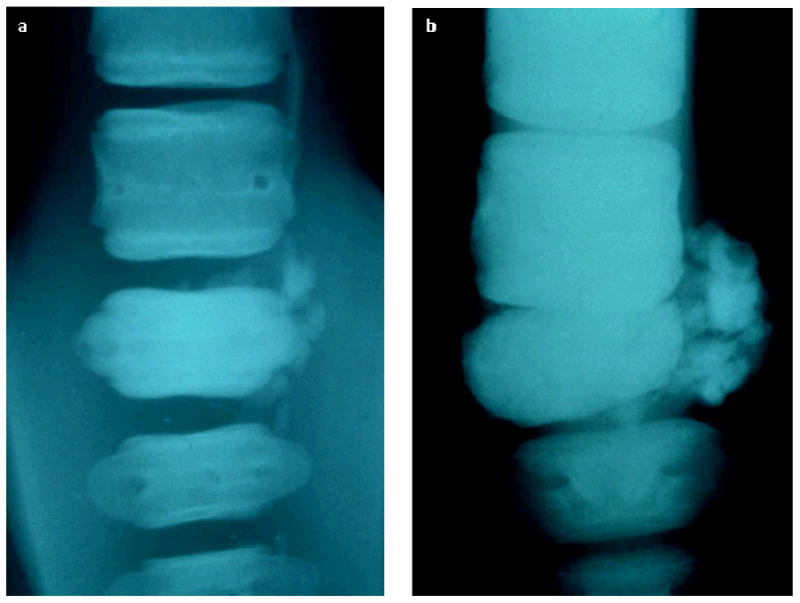
Radiographic imagings of the caudal vertebra taken at two different times, both images are a dorsoventral view with the more anterior vertebrae positioned at the top of the image. (a) Image taken shortly after arriving at Mystic Aquarium, showing sclerosis of the affected vertebra with soft-tissue calcification in the adjacent tissues but normal intervertebral spacing. (b) Image taken postmortem, 19 months later, showing further progression of the sclerosis of the affected vertebra and soft-tissue calcification as well as intervertebral chips, widening of the nutrient foramen, and collapse of the intervertebral space apparently allowing contact of the affected vertebra with the next most anterior vertebra