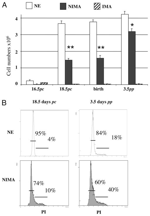
FIGURE 4.
Fetal and neonatal NIMA-exposed T cells are partially deleted from the thymus. Thymus T cells from NE or NIMA mice were analyzed by flow cytometry, using the Ti98 mAb specific for the BM3.3 Tg TCR. A, Compilation of cell numbers (± SEM) of BM3.3 Tg thymic T cells from NE control, NIMA, or IMA animals as 16.5- and 18.5-d pc fetuses, newborns, or 3.5-d-old neonates; at least seven animals were studied in each group in three separate experiments. *p,<0.05; **p,<0.01. B, Flow cytometry profiles obtained from NE or NIMA BM3.3 Tg thymic T cells from 18.5-d pc fetuses and 3.5-d-old neonates, after per-meabilization and staining with propidium iodide (PI). Percentages of cells in G0/G1 or S/G2/M phases of the cell cycle are given in each graph. One experiment representative of at least three for each group is shown.

