Figure 1.
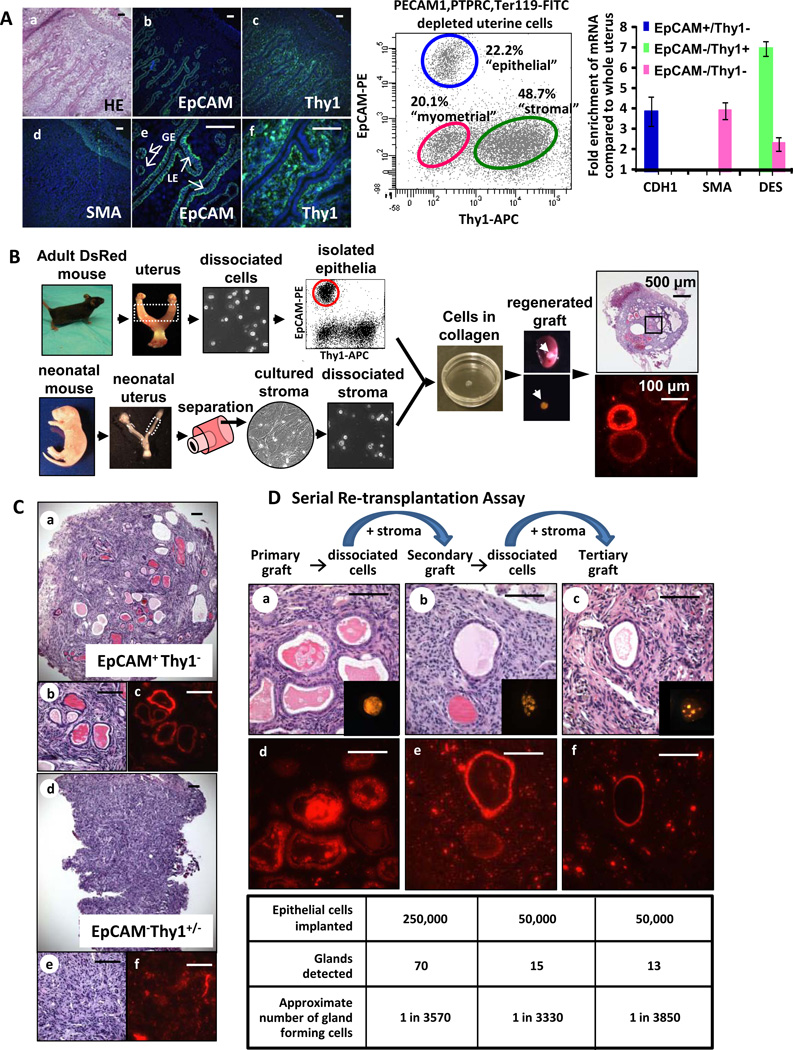
Adult mouse endometrial epithelia contain subpopulations of self-renewing cells. (A) Immunohistochemical staining showed that EpCAM marked all uterine epithelia: glandular epithelium (GE) and luminal epithelium (LE) (a,b&e). Thy1 marked the endometrial stroma (c&f) and smooth muscle actin (SMA) marked the myometrium (d). Epithelial (EpCAM+Thy1), stromal (EpCAMThy1+) and myometrial (EpCAMThy1) cells were isolated from dissociated uterine cellular preparations after exclusion of endothelial (PECAM1), lymphoid (PTPRC) and red blood cells (Ter 119). Quantitative PCR analysis confirmed identity of each cellular fraction based on enrichment for transcripts of E-cadherin (CDH1 epithelial), smooth muscle actin (SMA myometrial) and Desmin (DES stromal). (B) Combinations of FACS isolated total endometrial epithelia from adult DsRed mice and cultured neonatal stroma were implanted under the kidney capsule of an immunodeficient mouse. These cells regenerated into endometrial tissue. Epithelia in this regenerated tissue were marked with DsRed indicating their origin from isolated adult endometrial epithelia. (C) Only the EpCAM+Thy1 cells could regenerate in vivo into endometrial epithelial structures (a–c vs. d–f) measured by the formation of hollow, RFP positive glands (b&c vs. e&f). EpCAMThy1−/+ cells predominantly gave rise to stromal cells (e&f). (D) A sub-population of endometrial epithelia could serially re-transplant in vivo demonstrating the existence of a long-term self-renewing population of endometrial epithelia. Primary grafts (a&d), when dissociated and re-implanted with equal numbers of fresh stroma, gave rise to secondary grafts (b&e). Secondary grafts likewise gave rise to tertiary grafts (c&f). All scale bars are 100 µm and results are mean ± SD. Abbreviations: APC, allophycoerythrin; DES desmin; EpCAM, epithelial cell adhesion marker; GE, glandular epithelium; HE, hematoxylin and eosin; LE, luminal epithelium; PE, phycoerythrin; PECAM, platlet/endothelial cell adhesion marker; PTPRC, protein tyrosine phosphatase receptor C; and SMA, smooth muscle actin.

