Figure 2.
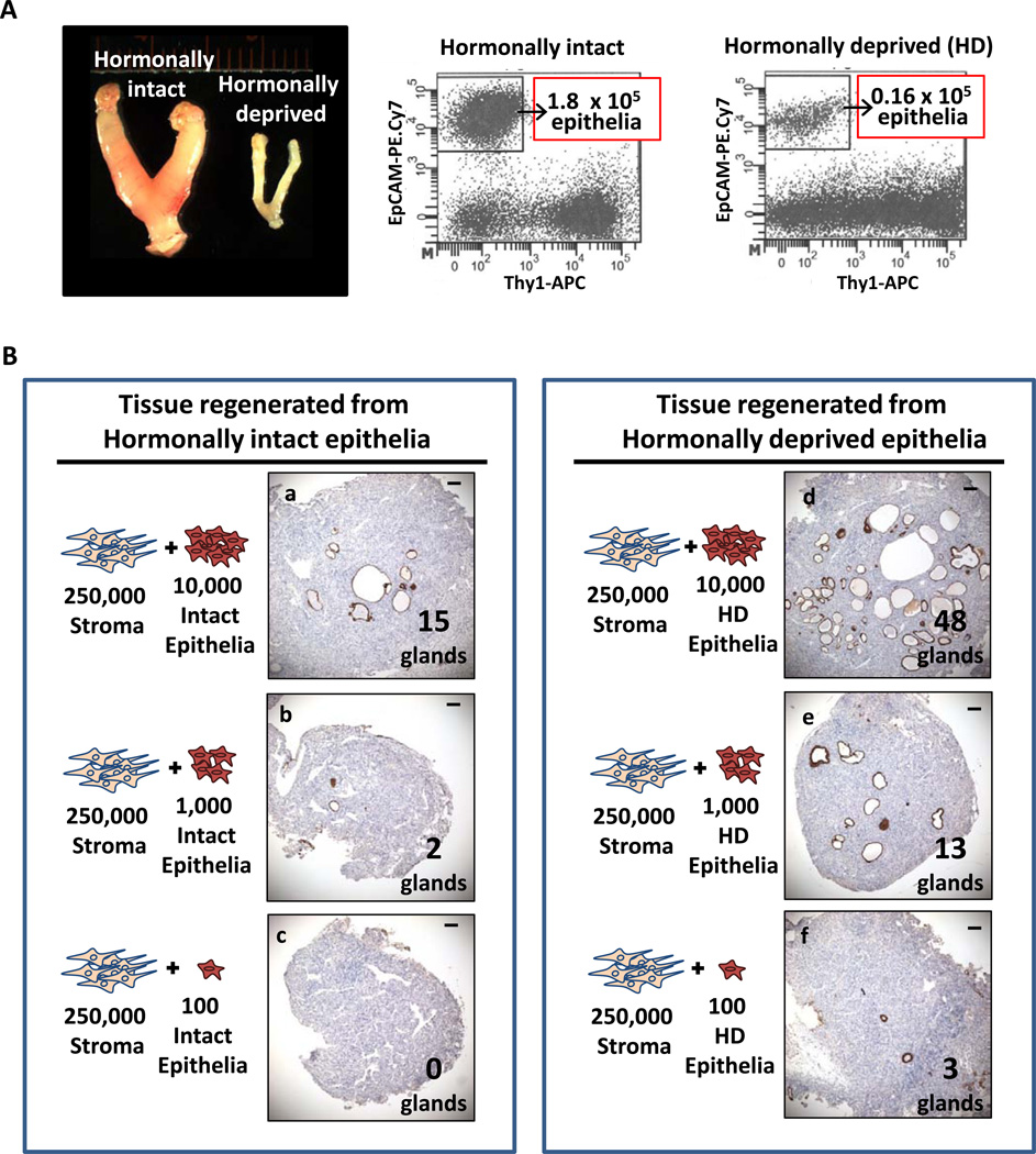
Hormonal deprivation resulted in a massive loss of endometrial epithelia but enrichment for endometrial epithelial progenitors. (A) Hormonal deprivation with surgical removal of the ovaries resulted in the shrinkage of the uterine horns and an approximate 10-fold drop in the number of total endometrial epithelia. (B) Endometrial epithelia that survived hormonal deprivation were enriched for epithelial progenitors. Equal numbers of endometrial epithelia from intact or hormone depleted uteri were placed in serial dilution in the in vivo regeneration assay. Regeneration was scored by the presence of pankeratin positive epithelial structures (glands). Representative regenerated grafts and the average number of glands detected per graft from the serial dilution series are shown. The in vivo growth capacity of hormonally deprived endometrial epithelia was superior to age matched hormonally intact cells at all serial dilutions tested. Scale bars equal 100 µm. Abbreviations: APC, allophycoerythrin; EpCAM, epithelial cell adhesion marker; HD, hormonally deprived; and PE, phycoerythrin.

