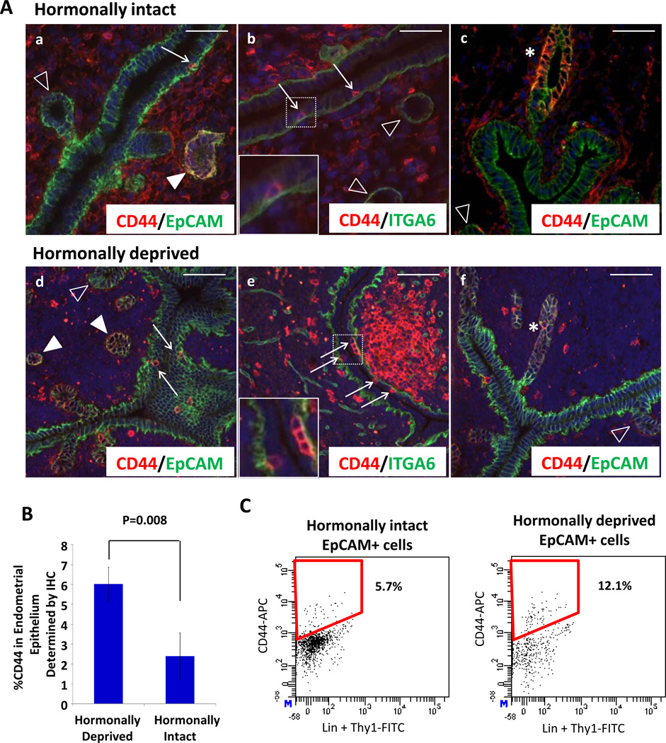
Figure 4.
Subsets of endometrial epithelia express CD44 and these cells are enriched in the hormonally deprived endometrium. (A) Isolated CD44 positive cells were seen in the epithelial lumen and in some but not all glandular epithelia (a–f). CD44 positive epithelial cells were in close contact with the basement membrane marked with ITGA6 (b&e). Arrows denote CD44 positive luminal epithelial cells (a,b,d&e). Filled arrow heads indicate CD44 positive epithelial glands (a&d). Empty arrow heads mark CD44 negative epithelial glands (b,c&f). Stars indicate positive luminal epithelial crypts (c&f). Boxed areas in b & e are shown as magnified insets. (B) An increase in the proportion of CD44 positive cells was detected upon hormonal deprivation in the endometrial epithelium. Based on immunohistochemistry, a higher percentage of CD44 positive epithelia were detected in hormonally deprived uteri. (C) FACS analysis also demonstrated an approximate doubling in the percentage of CD44 positive epithelia in the hormonally deprived endometrium compared to the hormonally intact conditions. All scale bars equal 50 µm and results are mean ± SD. Abbreviations: EpCAM, epithelial cell adhesion marker; FITC, fluorescein isothiocyanate.

