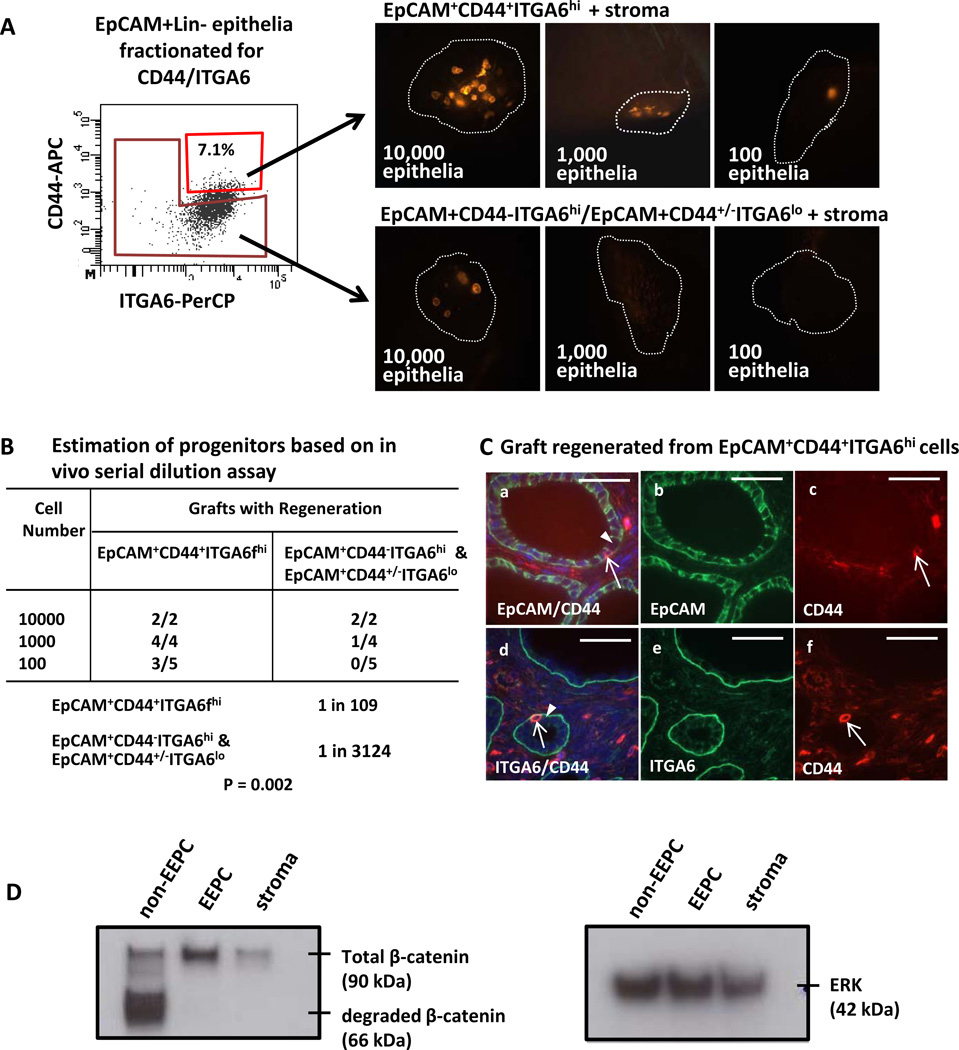
Figure 5.
Minor subpopulations of endometrial epithelia are EpCAM+CD44+ITGA6hiThy1−PECAM1−PTPRC−Ter119− and these cells are the endometrial epithelial progenitors (EEPC). (A) After depletion of Thy1+PECAM1+PTPRC+Ter119+ cells, endometrial epithelial were divided into two populations: (1) EpCAM+CD44+ITGA6hi, and (2) remaining endometrial epithelia comprised of EpCAM+CD44ITGA6hi and EpCAM+CD44+/−ITGA6lo cells. Logarithmic dilutions of equal numbers of these cells were grown in vivo. Regeneration was scored as the presence of RFP positive glandular structures. Predominance of the regenerative activity was detected in the EpCAM+CD44+ITGA6hi endometrial cells suggesting that these cells are the endometrial epithelial progenitors (EEPC). (B) A significant difference in the regenerative activity of these two subpopulations was detected in vivo based on a limiting dilution analysis. (C) EpCAM+CD44+ITGA6hi cells undergo multi-lineage differentiation in vivo. Majority of cells regenerated from this cellular pool were EpCAM/ITGA6 positive but CD44 negative (solid arrow heads) while a minor subpopulation of EpCAM/CD44 dual positive (a–c) and CD44/ITGA6 dual positive (d–f) cells were detected (arrow). (D) Higher levels of total β-catenin (90 kDa) were detected in EEPC vs. non-EEPC and stroma. Degraded β-catenin (66 kDa) was only detected in the non-EEPC. Scale bars equal 50 µm. . Abbreviations: APC, allophycoerythrin; EEPC, endometrial epithelial progenitor cell; and EpCAM, epithelial cell adhesion marker.

