
Fig. 3.
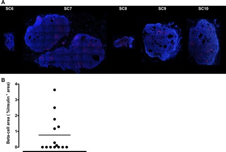
A: examples of epididymal pancreatic endodermal grafts extracted at 20 wk posttransplantation from nude rats. Representative examples of 6 epididymal pancreatic endodermal grafts stained by immunofluorescence for insulin (red) and nuclei (blue) at ×4 magnification taken from 5 different nude rats. B: quantification of insulin positive area from 14 different epididymal pancreatic endodermal grafts extracted from 5 nude rats after euthanization. Note that, in total, 3 grafts/animal (total of 15) were implanted at the beginning of the study, with 14 successfully retrieved following the completion of the study.