
Fig. 4.
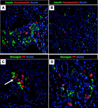
Examples of pancreatic endocrine cell expression in epididymal pancreatic endodermal grafts extracted at 20 wk posttransplantation from a nude rat with detectable human insulin/C-peptide levels. Representative images of epididymal pancreatic endodermal grafts stained for insulin/somatostatin (A and B) and glucagon/pancreatic polypeptide (PP; C and D) presented at ×20 magnification. Arrow denotes presence of glucagon and PP double-positive cells.