
Fig. 5.
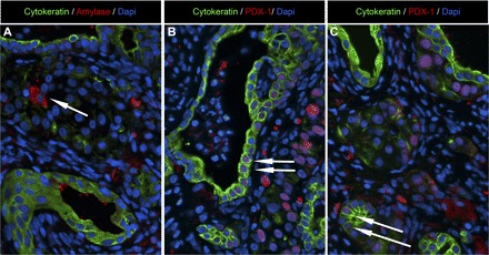
Examples of pancreatic exocrine cell expression in epididymal endodermal grafts extracted at 20 wk posttransplantation from a nude rat with detectable human insulin/C-peptide levels. A: representative image of epididymal pancreatic endodermal grafts stained for ductal cell marker (cytokeratin; green), exocrine cell marker (amylase; red), and nuclear marker DAPI (blue) presented at ×20 magnification. Arrow denotes presence of a single amylase positive cell. B and C: representative images of epididymal pancreatic endodermal grafts stained for ductal cell marker (cytokeratin; green), endocrine cell marker [pancreatic duodenal homeobox-1 (PDX-1); red], and nuclear marker DAPI (blue) presented at ×20 magnification. Arrows denote the presence of PDX-1/cytokeratin-positive cells throught the graft, including exocrine ductal tissue.