Fig. 6.

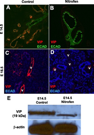
Immunofluorescent staining for VIP, a parasympathetic marker, in control and nitrofen-treated embryonic lungs. A and B: at E14.5, VIP is highly expressed within the distal airway epithelium, corresponding to nerve endings and/or neuroendocrine cells, in control embryonic lungs; this distal epithelial VIP expression is markedly reduced in nitrofen-treated embryonic lungs. C and D: at E16.5, VIP is also highly expressed within a perivascular pattern in control embryonic lungs; there is decreased perivascular VIP expression in nitrofen-treated embryonic lungs. V, blood vessels. Original magnification ×200. E: Western blot of VIP protein expression showing decreased VIP protein expression in nitrofen-treated embryonic lungs at E14.5 compared with control embryonic lungs at E14.5.
