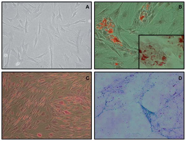
Figure 2.
(A) Fibroblast-like morphology of GTKO/hCD46 pAdMSC (light microscopy 10 ×). (B) Adipogenic differentiation of GTKO/ hCD46 pAdMSC: Oil Red staining (20 ×). (Insert: fat droplets stained with Oil Red, 40 ×). (C) Osteogenic differentiation of GTKO/ hCD46 pAdMSC: Von Kossa staining (20 ×), showing morphologic changes in pAdMSC and linear calcium deposition. (D) Chondrogenic differentiation of GTKO/hCD46 pAdMSC: Alcian Blue staining (10 ×), showing strongly acidic mucosubstance (blue) and goblet cells (red nuclei).

