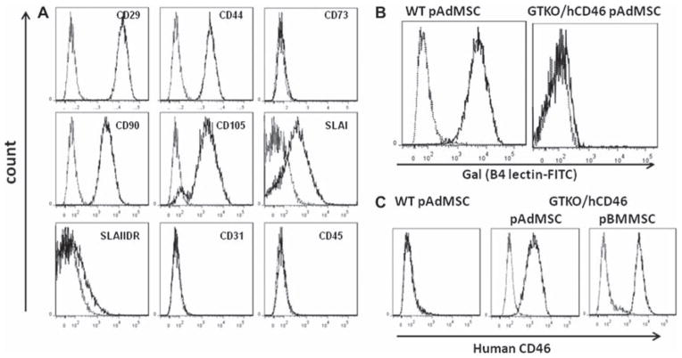
Figure 3.
(A) Flow cytometry characterization of GTKO/hCD46 pAdMSC, showing positive expression of CD29, CD44, CD90 and CD105, with negative expression of CD31, CD45, CD73 and SLAIIDR, and weakly positive expression of SLAI. (B) No surface expression of Gal was detected on GTKO/hCD46 pAdMSC compared with WT pAdMSC. (C) Expression of hCD46 on GTKO/hCD46 pAdMSC compared with expression on WT pAdMSC and on GTKO/hCD46 pBMMSC.

