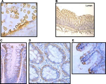
Fig. 3.

Immunolocalization of UT-B protein within human colon tissue using BUTB-PAN antibody (brown staining) and hematoxylin counterstaining (blue staining). A: BUTB-PAN stains plasma membrane of red blood cells within a human colonic artery (×100 magnification). B: longitudinal section (LS) of colon shows BUTB-PAN only stains the upper portion of the colonic crypts (×3 magnification). C: longitudinal section of colon shows BUTB-PAN signal is predominantly confined to the basolateral region of colonocytes situated in the upper crypt region, although some apical staining is also present (×63 magnification). D: transverse section (TS) of colon confirms BUTB-PAN signal is in the basolateral region of colonocytes (×63 magnification). E: longitudinal section of surface epithelial layer shows BUTB-PAN signal is present throughout the cytoplasm of these cells (×100 magnification).
