
Fig. 5.
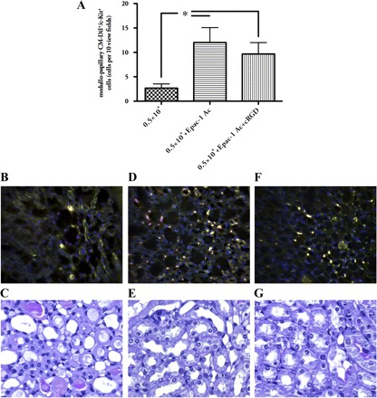
EPCs in postischemic kidneys. Animals injected with 0.5×10⁶ EPCs showed very sporadically EPC infiltrates in their respective kidneys (B). When cells pretreated with Epac-1 Ac alone were administered, renal c-Kit+/CM-Dil+ cell numbers were significantly elevated (D). Combined cell pretreatment with Epac-1 Ac and cRGD did not significantly change the number of double-positive cells in the postischemic organs (A and F). None of the analyzed kidneys showed any injected cells within the cortex. Most of the cells were located in the medullopapillary border zone. The images show representative cross sections of the papilla. A more detailed analysis of the cells' distribution is shown in Fig. 6. C, E, and G show representative areas of kidneys from the respective groups (PAS stained) [yellow: cells, double-positive for CellTracker (red) and FITC-c-Kit (green), magnification ×40 in B, D, and F, ×400 in C, E, and G; data are means ± SE, *P < 0.05].