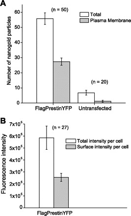
Fig. 3.

Equivalent amounts of prestin are found in the plasma membrane and cytoplasm. Quantitative analysis on the distribution of prestin molecules in a Flag-tagged prestin-YFP stable cell line is shown. A: nanogold particle counting from EM images. Individual particles were counted from each micrograph, and the particles on the plasma membrane were subtracted from the total. As shown here, on average there are 56 gold particles in each micrograph of transfected cells, out of which 27 are localized on the cell membrane (∼50.6 ± 2.5%). B: the intensity of fluorescence was assayed on Flag-tagged prestin-YFP-expressing cells. The localization of YFP fluorescence was quantified in the plasma membranes of these cells. The plasma membranes of these cells contained 48.6 ± 1.9% of the total fluorescence signal (± SE).
