
Fig. 1.
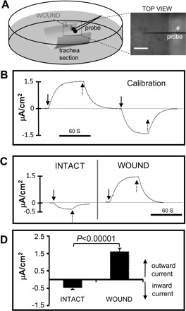
Wounds in trachea generate electric currents. A: schematic showing a hemi-cylindrical section of trachea mounted in a plastic 35-mm Petri dish containing BEGM culture medium. Trachea wound and vibrating probe were viewed under a dissection microscope (TOP VIEW, right). Scale bar = 0.5 mm. B: vibrating probe was calibrated in BEGM medium. After establishing a stable baseline (dashed line at 0), a current of 1.5 μA/cm2 was applied, first in one direction and then in the opposite direction. Solid arrows show when the current was switched on, and dashed arrows show when the current was switched off. Scale bar = 60 s. C: typical electric currents recorded from an intact and then wounded (wound was made immediately before the recording, <5 min prior) tracheal epithelium. Solid arrows show when the probe was brought to the measuring point, and dashed arrows show when it was brought away from the measuring point to the reference position. Inward/outward currents are below/above the baseline (dashed line at 0) respectively. Scale bar = 60 s. D: intact tracheal epithelium showed small inward currents. Small slit wounds had larger outward currents (P < 0.00001; n = 9).