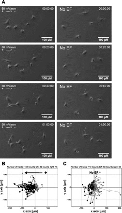
Fig. 3.

Direct current electrical field (EF) direct migration of tracheobronchial epithelial cells of rhesus monkey towards the cathode. A: cell movement in an EF of 50 mV/mm was recorded with time-lapse microscopy (left). Cells migrate randomly in control conditions without an EF (right). Each line with a number indicates an individual cell trajectory. Scale bar = 100 μm. B–C: migration trajectories. Each dot represents an individual cell with its trajectory plotted in a Cartesian coordinate system, in which the start point of each cell is set as the origin. Cells that migrate towards the right are highlighted in gray. In an EF cells migrate cathodally to the left (B), compared with the control (no EF) in which cells migrate randomly (C). EF = 50 mV/mm. Duration = 145 min. See Supplemental Files S1 and S2 for movies.
