
Fig. 6.
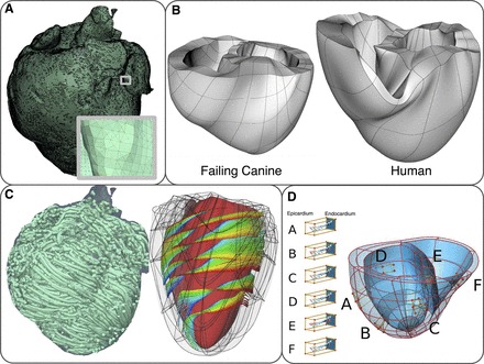
Computational meshes and fiber and sheet structure. A: computational mesh of infarcted canine ventricles for the electrical component of an electromechanics model. A, inset: zoomed-in image of the mesh, showing the mesh details. B: computational meshes for the mechanics component. C: fiber and sheet orientations obtained from diffusion tensor magnetic resonance images. Fiber orientation is for the infarcted canine heart. Sheet orientation is shown on the endocardium in diastole. D: human ventricles with rule-based fiber orientation. D, A–F: transmural changes in fiber angle for different regions in the ventricle. Figure and legend are modified from Trayanova et al. (227), used with permission.