
Fig. 7.
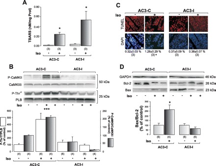
Mice with cardiomyocyte-delimited transgenic expression of CaMKII inhibitory peptide are protected from Iso-induced apoptosis. A: lipid peroxidation measured by TBARS indicates that Iso treatment increases oxidative stress in both AC3-C and AC3-I mice. B: representative blots from P-CaMKII and P-Thr17, and their respective total proteins, as an index of CaMKII activity, and average results, show that, under Iso treatment, only AC3-C mice increase phosphorylation of CaMKII and PLN at the CaMKII site. C: TUNEL and DAPI photographs of the different groups and mean (SE) values of these experiments, indicating a significant increment in TUNEL-positive cells normalized by total DAPI stained nuclei only in the AC3-C mice treated with Iso (***). D: typical blots of pro- and anti-apoptotic protein Bax, and Bcl-2, and mean (±SE) results in the bar graph below. GAPDH signals were used as loading controls. Iso treatment induced apoptosis only in AC3-C mice. *P < 0.05 vs. other groups. ***P < 0.001 vs. other groups.