
Fig. 1.
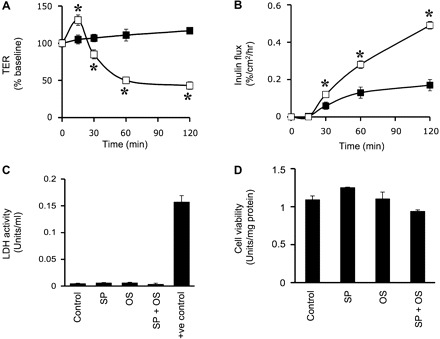
Osmotic stress (OS) disrupts tight junctions (TJs) in Caco-2 cell monolayers. A and B: Caco-2 cell monolayers were incubated with (□) or without (■) hyperosmotic buffer (600 mosM). Transepithelial electrical resistance (TER; A) and inulin permeability (B) were measured. Values are means ± SE (n = 6). *Significantly (P < 0.05) different from corresponding control values. C and D: Caco-2 cell monolayers were incubated with or without SP600125 (SP) for 1 h followed by exposure to OS for 1 h. C: aliquots of apical medium were assayed for lactate dehydrogenase (LDH) activity. D: the cells were assayed for cytotoxicity by WST-1 assay. Values are means ± SE (n = 6). Cells incubated with 0.1% Triton X-100 were used as a positive control.