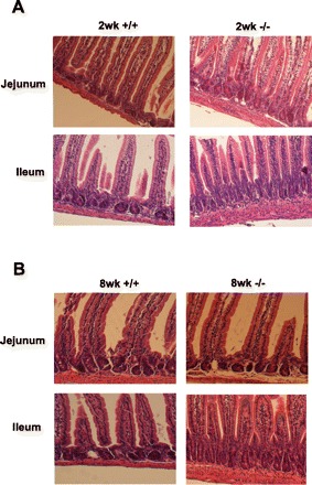
Fig. 3.

Morphological assessment of the small intestine. Small intestinal tissue was collected from male mice and fixed in 4% paraformaldehyde at 4°C overnight, dehydrated, and embedded in paraffin. Sections were stained with hematoxylin and observed under microscope (×100). +/+: Wild-type mice; (−/−): NHE8−/− mice. A: small intestine from 2-wk-old mice (2wk). B: small intestine from 8-wk-old mice (8wk).
