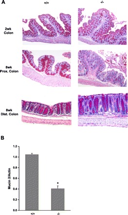
Fig. 6.

Mucin staining and MUC2 expression in the colon. A: large intestinal tissue was collected from male mice and fixed in 4% paraformaldehyde at 4°C overnight, dehydrated, and embedded in paraffin. Sections were stained with PAS and then observed under microscope (×100). +/+: wild-type mice; −/−: NHE8−/− mice. B: RNA was isolated from the colon and used for PCR analysis on MUC2 expression. MUC2 mRNA and β-actin mRNA were amplified with mouse-specific primers. Changes in MUC2 gene expression was analyzed by the semiquantitative method. Data are means ± SE from a total of 8 groups of mice (8- to 10-wk-old males and females). *P ≤ 0.01 for NHE8−/− mice (−/−) vs. wild-type mice (+/+).
