
Fig. 3.
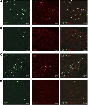
Confirmation of GFP expression in gastric oxyntic mucosal ghrelin cell. A: ∼90% of GFP cell (green) at the lower two-thirds of the gastric gland are ghrelin (Ghr)-immunoreactive (IR; red), and remaining GFP cells are not ghrelin-IR (arrow). GFP cells near the top surface of the gastric mucosa are not ghrelin-IR. B: GFP cells (green) do not colocalize with histidine decarboxylase (HDC)-immunopositive enterochromaffin-like cells (red). C: most GFP cells (green) are not somatostatin (Sst)-IR. Sst-IR cells (red) express a low green fluorescence (green, arrow) compared with Sst-negative GFP cells. D: GFP cells (green) do not colocalize with serotonin (5-HT)-producing enterochromaffin cells (red). All tissue sections for immunofluorescence staining are representative of multiple sections prepared from ≥3 mice.