
Fig. 4.
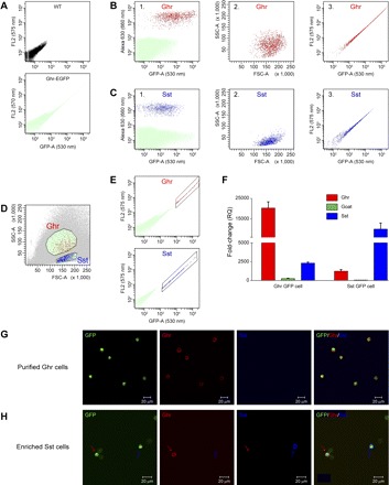
Flow cytometric analysis and isolation of gastric ghrelin-expressing GFP (Ghr-GFP) cells by fluorescence-associated cell sorting (FACS). A: fluorescence of dispersed gastric cells in wild-type (WT) mice and GFP cells in ghrelin-GFP transgenic mice. B: flow cytometric analysis of dispersed gastric cells immunostained for ghrelin. Presorted gastric cells from ghrelin-GFP transgenic mice were immunostained with anti-ghrelin antibody and Alexa Fluor 630-conjugated secondary antibody. B1: ghrelin-positive cells (red) were separated from non-ghrelin cells (green) on the basis of 630-nm emission. B2: ghrelin cells (red, identified in B1) were plotted on the basis of FACS forward scatter (FSC) and side scatter (SSC). B3: ghrelin-positive cells (red, identified from B1) were plotted on the basis of fluorescence intensity of GFP (530- and 575-nm emission). C: flow cytometric analysis of dispersed gastric cells immunostained for Sst. Presorted gastric cells from ghrelin-GFP transgenic mice were immunostained with anti-Sst antibody and Alexa Fluor 630-conjugated secondary antibody. C1: Sst-positive cells (blue) were separated from non-Sst cells (green) on the basis of 630-nm emission. C2: Sst cells (blue, identified in C1) were plotted on the basis of FSC and SSC. C3: Sst-positive cells (blue, identified from C1) were plotted on the basis of fluorescence intensity of GFP. D and E: FACS isolation of gastric Ghr-GFP cells. D: separation of ghrelin cells from Sst cells on the basis of SSC and FSC (B2 and C2). E: isolation of Ghr-GFP cells (red) and Sst-GFP cells (blue) on the basis of fluorescence intensity of GFP (B3 and C3). F: assessment of Ghr-GFP cell purification using quantitative RT-PCR of ghrelin, Sst, and ghrelin O-acyltransferase (GOAT). Transcript levels from isolated Ghr-GFP and Sst-GFP cells were compared with non-GFP cells. RQ, relative quantitation. G: demonstration that almost all isolated GFP cells (green) are Ghr-GFP cells. Parameters outlined in D and E were used to confirm that sorted cells were Ghr-GFP cells by specific immunoreactivity for ghrelin (red) and not Sst (blue). H: heterogeneity of Sst-GFP cells. Isolation of GFP cells using parameters for Sst cells, as outlined in D and E, resulted in only 30% Sst-IR (blue) and 15% ghrelin-IR (red) cells.