
Fig. 3.
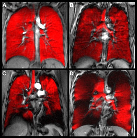
Hyperpolarized 3He-MRI of gas distribution (in red) coregistered to 1H-MRI of thoracic cavity (gray scale) of a healthy young never-smoker (A), older never-smoker (B), asthma (C), and COPD (D). MRI was obtained in the coronal plane during inspiration breath hold, after inhalation of a 1 liter gas mixture of hyperpolarized 3He and ultra-high purity N2 gas from functional residual capacity. 1H-MRI preceded 3He-MRI by ∼5 min and the 1H- and 3He-MRI slices were coregistered using rigid registration methods using the carina for fiducial landmarks. Coregistered 1H (gray scale)- and 3He-MRI gas distribution images show gas in red, and focal ventilation defects are clearly shown where the 1H thoracic cavity is exposed (in black) in the absence of gas. Note the absence of gas distribution abnormalities in A for the healthy young never-smokers, but there are qualitative differences for the elderly never-smoker and much more obvious abnormalities in asthma (C) and COPD (D). Some qualitative differences can also be observed in the gas distribution obvious in the trachea, although for all subjects, the breath-hold maneuver was the same.