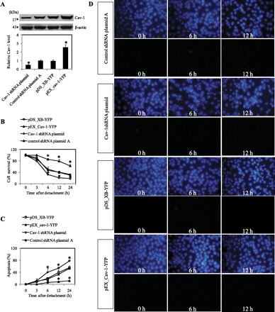
Fig. 1.

Caveolin-1 (Cav-1) overexpression increases anoikis resistance in H460 cells. A: control, HCav-1 [expression plasmid (pEX)_Cav-1 plasmid transfectant H460], or short hairpin (sh)Cav-1-transfected H460 cells were constructed and grown in culture that was then analyzed for Cav-1 expression by Western blotting. Blots were reprobed with β-actin antibody to confirm equal loading of samples. The immunoblot signals were quantified by densitometry, and mean data from independent experiments were normalized to the results. Columns are means ± SD (n = 3). *P < 0.05 vs. control transfected cells. B: subconfluent (90%) monolayers of control transfected, Cav-1-overexpressing cells, and Cav-1 knockdown cells were detached and suspended in poly-HEMA-coated plates for various times (0–24 h). At the indicated times after detachment, the cells were collected and their survival was determined by XTT assay. Viability of detached cells at time 0 was considered as 100%. C: percentage of cell detachment-induced apoptosis was analyzed by Hoechst 33342 nuclear fluorescence. Data points represent means ± SD (n = 3). *P < 0.05 vs. control transfected cells. D: detachment-induced apoptosis and necrosis in control transfected cells, Cav-1-overexpressing, and Cav-1 knockdown cells. Detached cells were suspended in poly-HEMA-coated plates for 0–12 h, and cell apoptosis and necrosis were determined by Hoechst 33342 and propidium iodide (PI) fluorescence measurements, respectively. pDS_XB-YFP, yellow fluorescent protein Cav-1 control plasmid.
