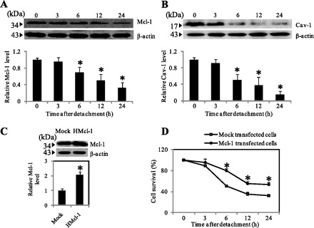
Fig. 2.

Cav-1 and myeloid cell leukemia sequence 1 (Mcl-1) expression after cell detachment. A and B: H460 cells were detached and suspended in poly-HEMA-coated plates for various times (0–24 h). Blots were probed with antibodies specific to Mcl-1 and Cav-1 and were reprobed with β-actin antibody. Columns are means ± SD (n = 3). *P < 0.05 vs. control at time 0. C: mock and HMcl-1 cells were grown in culture and analyzed for Mcl-1 expression by Western blotting. Blots were reprobed with β-actin antibody to confirm equal loading of samples. The immunoblot signals were quantified by densitometry, and mean data from independent experiments were normalized to the results. Columns are means ± SD (n = 3). *P < 0.05 vs. control transfected cells. D: subconfluent (90%) monolayers of Mock and HMcl-1 cells were detached and suspended in poly-HEMA-coated plates for various times (0–24 h). At the indicated times, the cells were collected and their survival was determined by XTT assay. Viability of detached cells at time 0 was considered as 100%. Data points represent means ± SD (n = 3). *P < 0.05 vs. control transfected cells.
