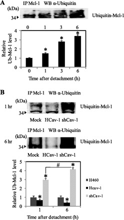
Fig. 5.

Effect of Cav-1 expression on Mcl-1 ubiquitination. A: H460 cells were detached and suspended in poly-HEMA-coated plates for various times. Cell lysates were prepared and immunoprecipitated (IP) with anti-Mcl-1 antibody. The resulting immune complexes were analyzed for ubiquitin by Western blotting (WB) using anti-ubiquitin antibody. Maximum Mcl-1 ubiquitination was observed at 6 h after cell detachment. The immunoblot signals were quantified by densitometry. Columns are means ± SD (n = 3). *P < 0.05 vs. control at detachment time = 0 h. B: HCav-1, shCav-1, and H460 cells were detached and suspended in poly-HEMA-coated plates for 1 and 6 h. Cell lysates were immunoprecipitated with anti-Mcl-1 antibody, and the resulting immune complexes were analyzed for ubiquitin (Ub) by Western blotting. Columns are means ± SD (n = 3). *P < 0.05 vs. control transfected at detachment time = 1 h; #P < 0.05 vs. the indicated control.
