
Fig. 5.
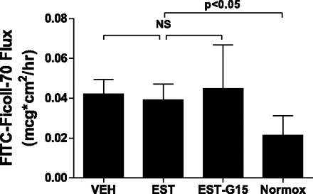
Transendothelial flux of FITC-Ficoll-70 after 8-h OGD and 8-h RGR. Cells were treated with vehicle, 100 nM 17β-estradiol, or 100 nM 17β-estradiol and 1,000 nM G15 (a selective GPR30 antagonist). Control cells were exposed to normoxic conditions in vehicle. Drug treatments were removed following OGD. At the 8-h time point, the protective effect of estrogen was lost. Drug-treated cells were not different from vehicle-treated cells, and all OGD-exposed cell monolayers transited more FITC-Ficoll-70 compared with control (Normox) cell monolayers. Values are means ± SD.