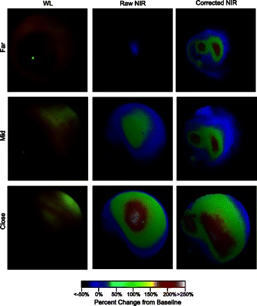
Fig. 3.

In vivo frames from mouse colonoscopy demonstrates quantitation of fluorescence signal in real time. WL, raw NIR, and corrected NIR images at different distances were acquired after orthotopic implantation of HT-29 colorectal cancer cells in the descending colon of a nu/nu mouse. Fluorescence colonoscopy was performed 24 h after intravenous injection of a protease-activatable optical probe. Raw NIR signal varied dramatically as the endoscope approached the target tumor; quantitative fluorescence intensity, however, was relatively invariant of the changes in distance between the tumor and catheter tip. Moreover, corrected NIR imaging was able to differentiate between 2 discrete tumor foci, whose presence was confirmed by subsequent histological analysis (data not shown). Adapted from Ref. 117.
