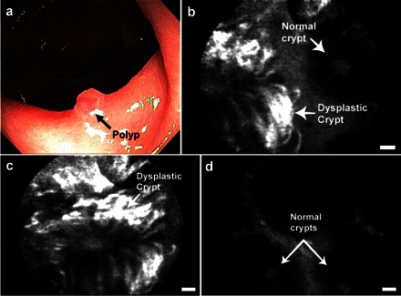
Fig. 5.

Optical molecular imaging of colorectal cancer in humans. a: white light imaging of a colon polyp by conventional colonoscopy. b: confocal microendoscopy following topical application of a fluorescently labeled peptide identified by phage display targeted to colorectal cancer identifies the border between normal and dysplastic tissue on a scale of micrometers. c: closer view of the dysplastic crypt shows increased binding of the labeled peptide compared with d, adjacent normal mucosa. Scale bars are 20 μm. Reproduced with permission from Ref. 53.
