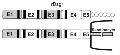
Figure 1.
The structure of desmoglein-1. Dsg1 contains 5 major cadherin-like domains on its extracellular portion. The black strips are the Ca2+ binding sites. The recombinant Dsg1 used in this study comprises the entire ectodomain of the molecule and is represented by the horizontal bar. There is a 6-histidine tail immediately downstream of the rDsg1, represented by circles.

