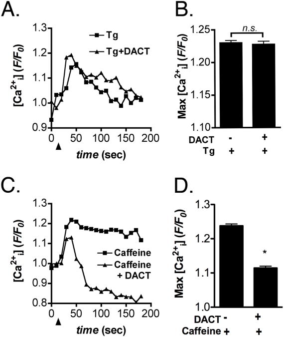
Figure 3. DACT selectively depletes ER calcium stores in LβT2 cells.
LβT2 cells primed by GnRH exposure were exposed for 24 hrs to 300 μM DACT or vehicle control (DMSO) and examined for changes in intracellular Ca2+ following stimulation with either 10 μM thapsigargin (Tg) or 5 mM caffeine. (A,B) Tg-induced release of Ca2+ from ER stores was unaffected in LβT2 cells treated with DACT, relative to vehicle-treated control cells. (C,D) Caffeine-induced release of Ca2+ from ryanodine receptor-sensitive stores was reduced following treatment with 300 μM DACT. Data were combined from 3 independent experiments, averaging 500 - 600 cells total per experimental group. (*p<0.05)

