
Fig. 6.
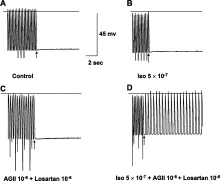
In vitro recordings from an excised Purkinje site of origin of focal VT labeled as in Fig. 4, including calibrations. A and B: no DAD/TA with pacing at 3 Hz in control or with addition of Iso, although spontaneous phase 4 depolarization is present, indicating Purkinje origin. C: after washout of Iso, addition of AGII and losartan superfusion did not result in DAD/TA, but phase 4 depolarization persists. D: with Iso, AGII, and losartan, pacing at the same frequency as A, B, and C now results in sustained TA.