
Fig. 7.
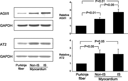
Western blot analysis of AGI/II and AGII receptor type 2 (AT2) protein expression in Purkinje fiber, and myocardium with and without ischemia (IS). Representative Western blots (left) correspond to graphs showing means ± SE (right). AGI/II and AT2 are expressed in both Purkinje fiber and cardiomyocytes. AGI/II and AT2 expression were higher in myocardium than those in Purkinje fibers. Ischemia significantly increased AGI/II expression, but not AT2, in cardiomyocytes. Expression levels of protein were normalized to those of GAPDH and are shown as fold increase over Purkinje fibers.