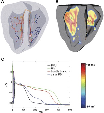
Fig. 1.

Computer model. A: three-dimensional model of rabbit ventricles with branching Purkinje system (PS). Colors indicate membrane potential (Vm; see the color bar). The same color scheme is used throughout the paper. B: excitation of the ventricles by the PS during normal sinus rhythm. Colors are as in A. C: action potentials (APs) in various parts of the conduction system. AP duration (APD) is longer in the bundle of His and bundle branches than in distal regions due to electrotonic interaction between terminal Purkinje cells and coupled myocytes. PMJ, Purkinje-myocardial junction.
