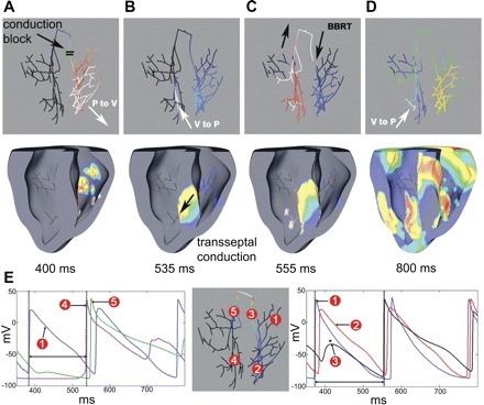
Fig. 6.

Bundle branch reentrant tachycardia (BBRT). A: ectopic activity spread through the right distal PS and into the RV (white arrow). CB was observed in the RBB. B: activations reached the LV by transseptal conduction (black arrow) and were picked up by the left PS 165 ms after the ectopic beat (white arrow). C: activations were conducted retrogradely through the LBB and bundle of His to reach the RBB after 185 ms. D: excitation spread through the right distal PS and RV. E: individual AP traces at various locations in the PS. Ectopic beat was originated near trace 1. *Retrograde CB in the RBB. The two-way arrow between dotted lines in the lefthand traces (between trace 1 and 4) indicate the time required by the ectopic beat to travel from right PS to the left PS (through septum). The two-way arrow between the dotted lines in the righthand traces (between trace 1 and 3) indicate the time taken by the ectopic activation to reach back to the RBB via LBB and His bundle.
