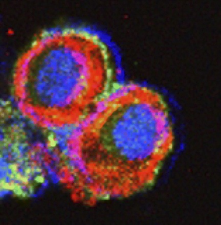
Fig. 4.

Immunocytochemical visualization of GABAA π receptor localization on confocal laser microscopy. Cell periphery was stained with Texas red, using an antibody against cytokeratin 5/6/18. GABAA π receptors were stained with fluorescein. The nucleus was stained with Topro-3. Cells were transfected with pcDNA-HOXA10, leading to greatly enhanced peripheral localization of the receptor.
